News
SAODS – Volume 5 Issue 11

| Publisher | : | Scienticon LLC |
|---|---|---|
| Article Inpress | : | Volume 5 Issue 11 – 2022 |
| ISSN | : | 2642-1623 |
| Issue Release Date | : | November 01, 2022 |
| Frequency | : | Monthly |
| Language | : | English |
| Format | : | Online |
| Review | : | Double Blinded Peer Review |
| : | editor@saods.net |
Volume 5 Issue 11
Research Article
Volume 5 | Issue 11
Abdelrahman Mustafa El sokkary, Noha El Khodary and Nermeen Nagi
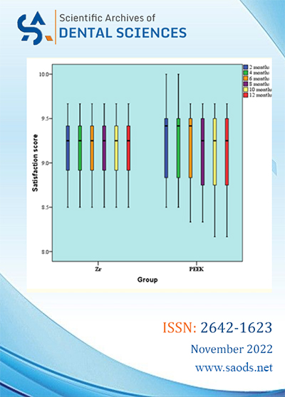
Aim: Comparing the clinical performance of CAD/CAM BioHPP PEEK- based crowns to zirconia veneered crowns through evaluation of patient satisfaction.
Methodology: 24 full coverage crowns were fabricated for Molars. Scaling and polishing were performed for all the patients one week prior to preparation. Regarding crowns’ material patients were divided into 2 main groups: In Group 1 (control group) patients received Zr veneered crowns while in Group 2 (intervention group) patients received PEEK crowns. Supra- gingival, chamfer finish line is performed for all teeth during preparation as a method of standardization. CAD/CAM (CAM5-S1) machine with software (Exocad) was used for performing try-in and provisionalization. All restorations were veneered and surface treated regarding the manufacturer’s instructions. All the crowns were cemented by using self-adhesive resin cement (by BISCO). During follow up visits evaluation of shade and function to determine patient satisfaction by questionnaire. Measurements were repeated every (2- 4- 6- 8-10 and 12 months respectively.
Results: Fisher’s Exact test was used in comparison between groups. There was no statistically significant difference between the 2 groups (P-value = 1, Effect size = 0.478) for every time.
Conclusion: Both PEEK crowns and Zr veneered crowns revealed successful clinical performance from patient satisfaction and clinical performance aspect. The two materials showed no significant difference; regarding the patient satisfaction and clinical performance.
Keywords: Patient Satisfaction; Biocompatibility; Zirconium Veneered Crowns; PEEK Crowns
Case Report
Volume 5 | Issue 11
Marcelo Yoshimoto, Jefferson de Oliveira Carvalho, Jefferson Pereira Xavier, Zilma de Oliveira Souza, Marcelo do Lago Pimentel Maia and Irineu Gregnanin Pedron
The loss of bone volume after exodontia in the posterior region of the mandible can be caused by several reasons. In childhood, early loss may occur due to caries and trauma. In adulthood, tooth loss can be caused by caries, trauma, periapical pathology, periodontal disease or traumatically performed extractions. The loss of the bone structure supporting the implants leads to another problem. In the posterior region of the mandible, the installation implants is made more difficult by the presence of the inferior alveolar nerve. To meet this need, some possibilities of treatment are presented, such as bone block grafts, short implants or inferior alveolar nerve lateralization surgery. The purpose of this article is to compare six cases in which short implants and inferior alveolar nerve lateralization surgery were performed for implant installation.
Keywords: Short Implants; Inferior Alveolar Nerve Lateralization Surgery; Mandibular Bone Atrophy; Injury to the Inferior Alveolar Nerve
Case Report
Volume 5 | Issue 11
Denis Jim Palomino Gutiérrez, Henry Jeancarlo Obispo Ruiz, Jose Luis Flores Mesia, Aldair Alfredt Lopez Malaga, Aurelio Medina Gonzáles, Israel Ribeyro Vásquez and María Angélica Fry Oropeza
Diabetic patients have to be treated with a prevention protocol since they are susceptible to infections.
Different preventive measures are used such as: premedication, mouthwashes, scaling and root planing, prophylaxis, defocusing (exodontia of pieces with loss of support and periodontal insertion), karyological treatment, oral rehabilitation. Among the delocalization protocol in diabetic patients, where extractions are to be performed, it is established as a rule after the evaluation to perform a panoramic radiograph, eventually a periapical radiograph (when you want to evaluate a sign located in the piece). Degree of infection in the tooth and type of premedication, elimination of predisposing factors of periodontal disease through prophylaxis, elimination of the stone. Despite all the preventive protocols, there is always a latent condition of complications, for this reason publications have been found in different scientific articles on the use of ozone gas as a complementary element in disinfection of oral pathogens, oxygenation of tissues and a beneficial effect on healing.
With this objective, we found an innovative and complementary protocol to reduce the bacterial load before dental extraction with ozone gas topication at the level of the gingival margin and papillae, both at the level of the preoperative preparation (prophylaxis, scaling, scaling and root planing). as at the time before the extraction, and once the extraction, coagulation formation and ozonation of the surface and gingival mucosa of the tooth extracted, a success rate greater than 50% is expected, compared to the protein structures of the pathogens.
In the following work, we are going to address the issue of medical ozone applied in dentistry, in a controlled patient with diabetes, for which the concentration of medical ozone in % 30Ug was used to help reduce the bacterial load, due to the properties that it has this gas against oral tissues and against pathogens, in this case of greater relevance, such as an insulin-dependent diabetic patient.
Keywords: Medical Ozone; Surgery; Protocols; Teeth; Disinfection; Tooth Extraction; Diabetes; Cicatrization; Oxidation; Oral Hygiene Index
Commentary
Volume 5 | Issue 11
Danilo Lourenço and Irineu Gregnanin Pedron
Opinion
Volume 5 | Issue 11
Irineu Gregnanin Pedron